


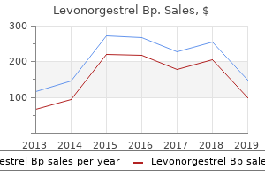
"Generic 0.18 mg levonorgestrel amex, birth control lo loestrin fe".
By: Z. Gamal, M.A., M.D., M.P.H.
Assistant Professor, University of Illinois at Urbana-Champaign Carle Illinois College of Medicine
Portions of the coracohumeral ligament type a tunnel for the biceps tendon on the anterior aspect of the joint birth control pills libido order 0.18mg levonorgestrel with amex. It converts the inter tubercular sulcus into a canal birth control 6 months shot levonorgestrel 0.18 mg amex, and acts as a retinaculum for the lengthy tendon of biceps birth control quick start order 0.18mg levonorgestrel with visa. Synovial membrane the synovial membrane traces the capsule and covers components of the anatomical neck. The intraarticular bicipital sheath may be linked to the articular floor of the superior capsule above by a mesotenon, which can be incomplete, presenting as one or a quantity of strands. This arrangement can only be the case if the biceps tendon was originally an extraarticular structure and invaginated, with its synovial sheath, into the glenohumeral joint because the upper limb rotated into external rotation relative to the trunk during growth. They are often discovered between the tendon of subscapularis and the capsule, speaking with the joint between the superior and middle gleno humeral ligaments; on the superior acromial aspect; between the cora coid course of and capsule; between teres major and the lengthy head of triceps; and anterior and posterior to the tendon of latissimus dorsi. Bursae typically happen behind coracobrachialis and between the tendon of infraspinatus and the capsule, often opening into the joint. The superior glenohumeral ligament passes from the sup raglenoid tubercle, simply anterior to the origin of the lengthy head of biceps, to the humerus at the fovea capitis, close to the proximal tip of the lesser tubercle on the medial ridge of the intertubercular sulcus. It types an anterior cover around the lengthy head of biceps and is a part of the rotator interval. Together with the coracohumeral ligament, it is an important stabilizer within the inferior direction, helping to keep the humeral head suspended (the coracohumeral ligament is extra sturdy than the tremendous ior glenohumeral ligament). The center glenohumeral ligament arises from a large attachment beneath the superior glenohumeral ligament, along the anterior glenoid margin as far as the inferior third of the rim, and passes obliquely inferolaterally, enlarging because it does so, to connect to the lesser tubercle deep to the tendon of subscapularis, with which it blends. The width and thickness of this ligament could additionally be as a lot as 2 cm and four mm, respectively. It provides anterior stability between 45� and 60� of abduction within the scapular plane. It could additionally be thickened and cordlike within the Buford advanced (a congenital glenoidal labrum variant), again with no apparent drawback for stability. The thicker and longer inferior glenohumeral ligament complex is a hammocklike structure with anchor points on the anterior and pos terior sides of the glenoid. It arises from the anterior, center and posterior margins of the glenoid labrum, beneath the epiphysial line, and passes anteroinferiorly to the inferior and medial features of the neck of the humerus. Flexion� extension, abduction�adduction, and medial (internal) and lateral (external) rotation all happen at the shoulder. Although the majority of the movement of the shoulder occurs on the glenohumeral joint, the scapulothoracic articulation contributes to overall shoulder movement in all directions, including lateral rotation. Muscles producing movements the muscles that produce transfer ments on the glenohumeral joint are principally the scapulohumeral and thoracobrachial muscular tissues: deltoid, pectoralis major and latissimus dorsi, assisted by coracobrachialis. The translating impact of those muscle tissue on the shoulder joint is counteracted by the rotator cuff, a group of brief muscular tissues (subscapularis, supraspinatus, infraspinatus and teres minor), together with teres main, that are connected closer to the joint, and that centre the top of the humerus in the glenoid fossa via the complete range of movement. Flexion Flexion is carried out by pectoralis major (clavicular part), deltoid (anterior fibres) and coracobrachialis, assisted by biceps. The capsule is equipped by the suprascapular nerve (posterior and supe rior parts), axillary nerve (anteroinferior) and the lateral pectoral nerve (anterosuperior, together with the rotator interval capsule). However, in clinical follow, actions are associated to the traditional anatomical planes. When the arm hangs at rest, the glenoid fossa faces almost equally forwards and laterally, and the humeral capitular and scapular (topographical) axes correspond, although the humerus, relative to the anatomical place, is medially rotated. Using the anatomical description, flexion carries the arm antero medially on an axis through the humeral head orthogonal to the glenoid fossa at its centre. Abduction and adduction occur in a vertical aircraft orthogonal to that of flexion�extension and the axis is horizontal, via the humeral head and parallel with the glenoid plane. However, when referred to the trunk, flexion and extension occur in the paramedian aircraft, and abduction and adduction occur in the coronal aircraft. In this sense, raising the arm vertically from flexion or raising it from abduction are each accompanied by humeral rotation in opposite directions. Abduction might reach 180� after thoracoscapular arthrod esis for facioscapulohumeral dystrophy (Copeland and Howard 1978). Some 60� of further abduction happens on the sterno and acromiocla vicular articulations, and contralateral vertebral flexion additionally aids in bringing the arm to the vertical.
If a compartment syndrome is suspected birth control pills ivf purchase 0.18 mg levonorgestrel amex, compartment stress measurements could additionally be indicated birth control guide order 0.18mg levonorgestrel overnight delivery, however birth control for women 6 months cheap levonorgestrel 0.18mg on-line, in unequivocal instances, the definitive treatment is emergency fasciotomies of the forearm. The absolute intracompartmental pressure threshold for decompression is the subject of debate: an absolute value of 30 mmHg is mostly accepted, although some authorities prefer a determine that comes inside 30 mmHg of diastolic pressure. Incisions to decompress the flexor compartment (Ronel et al 2004) run from the medial epicondyle and curve down the anterior aspect of the forearm, ending on the radial aspect of flexor carpi ulnaris; from right here, they could be prolonged distally to decompress the carpal tunnel, if essential. The bicipital aponeurosis (lacertus fibrosus) is routinely launched to decompress the median nerve. After retraction of the superficial flexors, the fascia over the deep muscular tissues of the forearm may be incised over its full length. Both median and ulnar nerves should be inspected to make positive that their decompression is full. The mobile wad and dorsal compartment are decompressed by way of a straight longitudinal incision that starts just distal to the lateral epicondyle; that is deepened through the airplane between extensor digitorum and extensor carpi radialis brevis, and ends over the midline of the wrist. In both cases, the injuries are left open and dressed, and only closed when circumstances allow. Pronator teres is medial to the proximal part of the artery, and the tendon of flexor carpi radialis is medial to the distal portion. The superficial radial nerve lies lateral to the center third of the radial artery; multiple branches from the artery provide the nerve all through its size. The radial artery could occasionally arise from a continuation of a superficial brachial artery, or as a excessive proximal division of an otherwise regular brachial artery. It passes between the superficial radial nerve and the posterior interosseous nerve earlier than ascending beneath brachioradialis, anterior to supinator and brachialis. It supplies these muscles and then anastomoses with the radial collateral department of the profunda brachii artery. The radial recurrent artery also offers branches to extensor carpi radialis longus, extensor digitorum, extensor digiti minimi and extensor carpi ulnaris. Ulnar collateral ligament Extensor carpi ulnaris (cut tendon) Anconeus (cut tendon) Anular ligament Interosseous recurrent artery Supinator Anconeus Posterior interosseous nerve Extensor carpi ulnaris Flexor carpi ulnaris Flexor digitorum superficialis Posterior ulnar recurrent artery Flexor digitorum profundus Muscular branches Muscular branches are distributed to the muscular tissues on the radial side of the forearm. Direct branches from the radial artery provide the radial insertion of pronator teres, the anterolateral aspect of flexor carpi radialis, the lateral surface of flexor digitorum superficialis, the lateral half of flexor pollicis longus, and the distal sections of extensor carpi radialis longus and extensor carpi radialis brevis. It is bigger than the radial artery and is accompanied all through its length by venae comitantes. In the forearm, the ulnar artery initially lies on brachialis before passing deep to pronator teres, flexor carpi radialis, palmaris longus and flexor digitorum superficialis. It then lies on flexor digitorum profundus, between flexor carpi ulnaris and flexor digitorum superficialis, and is roofed by the skin and superficial and deep fasciae. The artery crosses the flexor retinaculum, lateral to the ulnar nerve and pisiform bone, to enter the hand. The ulnar nerve lies medial to the distal two-thirds of the artery, which supplies the nerve throughout its length; the palmar cutaneous branch of the ulnar nerve descends alongside the ulnar artery to reach the hand. The ulnar artery may come up above the elbow, when it can lie superficial to the forearm flexors beneath the deep fascia; solely not often is it subcutaneous. When this happens, the brachial artery provides the common interosseous and the ulnar recurrent arteries. It is overlapped by contiguous sides of flexor digitorum profundus and flexor pollicis longus. This accompanies and supplies the median nerve so far as the palm, where it might be a part of the superficial palmar arch or finish as one or two palmar digital arteries. The median artery can even arise from the ulnar or the widespread interosseous artery. Muscular and nutrient branches from the anterior interosseous artery pierce the interosseous membrane to provide deep extensor muscular tissues and the radius and ulna, respectively. The anterior interosseous artery leaves the anterior compartment by piercing the interosseous membrane proximal to pronator quadratus. It anastomoses with the posterior interosseous artery within the posterior compartment of the forearm, and travels via a tunnel beneath the extensor retinaculum with the tendons of the digital extensors before joining the dorsal carpal arch.
Buy levonorgestrel in india. 3 Completely Unknown Side Effects of Birth Control Pills.
The ascending lumbar vein is variable in its course and connections; hardly ever birth control pills that cause weight loss purchase levonorgestrel on line amex, the entire vein or a phase could additionally be absent on one facet (Lolis et al 2011) birth control weight gain safe levonorgestrel 0.18 mg. It generally joins the subcostal vein to kind the azygos vein on the best and the hemiazygos on the left birth control pills questions and answers buy cheap levonorgestrel 0.18 mg on line. The azygos and hemiazygos veins run forwards over the twelfth thoracic vertebral body, and cross deep to or through the right and left crus of the diaphragm, respectively, into the thorax (Ch. The ascending lumbar vein is usually joined by a small vein, the lumbar azygos vein, from the back of the inferior vena cava or left renal vein. Sometimes, the ascending lumbar vein ends within the first lumbar vein, which then joins the lumbar azygos vein on the stage of the first lumbar vertebra. Blood circulate within the ascending lumbar veins can happen in both direction (Morita et al 2007). These branches anastomose posteriorly with tributaries of the azygos and hemiazygos veins, and anteriorly with branches of the epigastric, circumflex iliac and lateral thoracic veins. These superficial anastomoses present alternative routes of venous drainage from the pelvis and lower limbs to the center in the presence of inferior vena caval obstruction. The lumbar veins also communicate with the external and inside vertebral venous plexuses, offering an additional collateral pathway for venous return. The third and fourth lumbar veins usually cross forwards on the edges of the corresponding vertebral bodies to enter the posterior facet of the inferior vena cava; the left lumbar veins move behind the abdominal aorta and are, subsequently, longer. The first and second lumbar veins are rather more variable; they may drain into the inferior vena cava, ascending lumbar, lumbar azygos and renal vein (on the left), and are often linked to each other. Indeed, the first lumbar vein typically passes inferiorly to be part of the second lumbar vein or, less commonly, drains instantly into the ascending lumbar vein or passes forwards over the L1 vertebral body to be part of the lumbar azygos vein. The second lumbar vein may join the inferior vena cava at or near the extent of the renal veins or, much less commonly, joins the third lumbar or ascending lumbar vein. Both veins could additionally be changed by multiple vessels within the decrease stomach and generally remain double so far as their termination. Renal veins the renal veins are large-calibre vessels, which lie anterior to the renal arteries and open into the inferior vena cava nearly at proper angles (Ch. The left vein lies on the posterior belly wall posterior to the splenic vein and body of the pancreas. It passes to the right in the angle between the abdominal aorta posteriorly and the superior mesenteric artery anteriorly, to empty into the inferior vena cava. The proper renal vein lies posterior to the second part of the duodenum and, sometimes, the lateral part of the pinnacle of the pancreas. Lymph from the ovary can also drain via lymphatics within the broad ligament and spherical ligament to internal iliac and inguinal lymph nodes, respectively. Lymphatic drainage of the ureters is less well outlined but follows a regional pattern to nearby nodes. The lymphatic drainage of the retroperitoneal colon and rectum is described in Chapter sixty six. Whilst the clinical sample of nodal metastases often reflects regular lymphatic drainage pathways, lymphatic obstruction might result in metastases at uncommon nodal sites. Cisterna chyli and stomach lymph trunks the abdominal origin of the thoracic duct normally lies to the right of the midline on the level of the twelfth thoracic vertebral physique or the thoracolumbar intervertebral disc. It receives virtually all the lymph from below the diaphragm via the cisterna chyli, a localized lymphatic dilation shaped mostly by the union of the intestinal lymph trunk and the left lumbar trunk (Phang et al 2014). The cisterna chyli is a saccular or fusiform lymphatic dilation measuring, on common, about 1 cm wide and a pair of cm lengthy in the cadaver (Loukas et al 2007b). The cisterna chyli normally lies in entrance of the first and second lumbar vertebrae behind the right crus of the diaphragm to the best of the abdominal aorta but may be positioned at the next vertebral level. The most common configuration, found in roughly two-thirds of people, is a single construction formed from the union of the intestinal lymph trunk and the left (or, much less generally, the right) lumbar lymph trunk (Loukas et al 2007b). The higher two proper lumbar arteries and the right lumbar azygos vein lie between the cisterna chyli and the vertebral column. The medial edge of the proper crus of the diaphragm lies anterior to the abdominal confluence of lymph trunks.
Postganglionic sympathetic neurones from each sources launch noradrenaline (norepinephrine) birth control pills effectiveness discount levonorgestrel 0.18 mg line, causing presyn aptic inhibition within enteric circuits birth control migraines purchase 0.18mg levonorgestrel with mastercard, slowing gut motility and driving contraction of the ileocaecal and internal anal sphincters birth control pills yahoo buy 0.18mg levonorgestrel otc. Sympathetic provide to the midgut is conveyed to the coeliac and superior mesenteric plexuses via the larger and lesser splanchnic nerves; postganglionic axons are distributed with branches of the superior mesenteric artery. Sympathetic provide of the hindgut is conveyed by way of the lumbar splanch nic nerves that synapse in the abdominal aortic and inferior mesenteric plexuses, and by way of sacral splanchnic nerves that synapse within the superior and inferior hypogastric plexuses; postganglionic fibres are distributed with branches of the inferior mesenteric artery and are inhibitory to colonic muscle. The midgut receives its parasympathetic innervation from the vagus, via the coeliac and superior mesenteric plexuses, whereas the hindgut receives its parasympathetic innervation from the pelvic splanchnic nerves. The cell our bodies of the pelvic splanchnic nerves are positioned within the second to fourth sacral spinal segments, the sacral parasympathetic nucleus. These parasympathetic fibres enter the inferior hypogastric plexus, the place some synapse. From right here, some cross directly to the rectum and other pelvic viscera whereas others ascend by considered one of two routes: either throughout the hypogastric nerves to the superior hypogastric plexus to be distributed along branches of the inferior mesenteric artery, or by passing instantly through the retroperitoneal tissues to reach the splenic flexure and descending and sigmoid colon. Most preganglionic parasympathetic neurones synapse in intramural plexuses in the intestine wall; from here, postganglionic neurones innervate the glands (secreto motor) and muscle (motor) of the large intestine. Parasympathetic stimulation is integral to colonic propulsion and defecation and to relaxation of the interior anal sphincter. Visceral afferent impulses mediating sensations of distension and spasm from the midgut journey with the vagus nerve while the hindgut is innervated by afferent neurones with cell bodies within the lumbar (mostly L2 and L3) and sacral dorsal root ganglia (mostly S1 and S2) (Brookes et al 2009). Visceral afferent innervation of intramural colonic blood vessels probably conveys the feeling of colonic distension (Song et al 2009), while visceral afferents in the rectum convey the feeling of rectal filling and are concerned in reflex propulsive activity. The anal canal lies 2�3 cm anterior and slightly inferior to the tip of the coccyx, opposite the apex of the prostate in males. At the anal verge, the squamous epithelium lining the lower anal canal turns into continuous with the pores and skin of the perineum. The space of pigmentation of skin around the anal verge corresponds approxi mately to the extent of the exterior anal sphincter. The anal canal consists of an inside epithelial lining, a vascular subepithelium, the interior and exterior anal sphincters, and fibro muscular supporting tissue, as well as dense neuronal networks of autonomic and somatic origin. It is between 2 and 5 cm long in adults; the anterior wall is slightly shorter than the posterior. At rest, it forms an oval or triradiate slit within the anteroposte rior airplane rather than a very round canal. The association of the exterior anal sphincter and its attachments to the perineal body and coccyx create sites of most stress in the anterior and posterior midline of the canal. Anteriorly, the middle third of the anal canal is hooked up by dense connective tissue to the perineal physique, which separates it from the membranous urethra in males and from the lower vagina in females. Laterally and posteriorly, the anal canal is surrounded by the loose adipose tissue of the ischioanal fossae; this association permits expan sion of the canal but presents a potential pathway for the spread of peri anal sepsis. Posteriorly, the anal canal is connected to the coccyx through the anococcygeal ligament, a midline fibroelastic construction that runs between the posterior side of the middle area of the exterior anal sphincter and the coccyx. The anococcygeal ligament is historically considered lying just inferior to the midline raphe of levator ani however its relationship to the raphe is more complicated (Kinugasa et al 2011). The ischial spines could additionally be palpated laterally by an analyzing finger in the upper anal canal. The pudendal nerves cross over the attachment of the sacrospinous ligament at this level and pudendal nerve motor terminal latency could also be measured digitally using a modified electrode worn on the examining glove. The subepithelial tissues are cellular and relatively distensible, and comprise submucosal arterial and venous plexuses. The columns frequently contain a terminal branch of the superior rectal artery and vein, supplemented to a variable degree by middle and infe rior rectal vessels (Thomson 1975). Dilated submucosal veins in the upper anal canal type an inner haemorrhoidal venous plexus. Tiny arteriovenous connections to these dilated submucosal veins give the blood within them a higher oxygen tension and due to this fact a redder color than regular venous blood. Although variable in quantity and place, the cushions help to seal the anal canal and contribute to the upkeep of continence to flatus and fluid. The lower ends of the columns form small crescentic folds, referred to as anal valves, between which lie small recesses known as anal sinuses.