


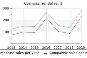
"Cheap 5 mg compazine visa, medicine video".
By: I. Marlo, M.B. B.CH., M.B.B.Ch., Ph.D.
Assistant Professor, A.T. Still University School of Osteopathic Medicine in Arizona
The second cervical nerve exits from the cervical canal immediately adjacent and dorsal to the joint capsules treatment trichomoniasis buy generic compazine on line. The transverse atlantal ligament is a band three to five mm thick that originates from the tubercles and the inside facet of the lateral masses of the atlas vertebra and is in close apposition to the odontoid; this ligament permits axial rotation symptoms ketoacidosis purchase 5 mg compazine overnight delivery. By itself, the geometry of the craniovertebral complicated is meant to provide mobility at the value of stability medicine 3 sixes buy compazine 5mg on-line. The frequent prevalence of patterns with various combinations suggests an interrelationship between if not a common reason for the origin and improvement of these constructions. The majority of the skull and facial bones develop by intramembranous ossification. Such growth bypasses the intermediate cartilaginous stage attribute of growth of the bony cranial base. The third sclerotome is responsible for the exoccipital middle as it types the jugular tubercles. The hypocentrum of the fourth occipital sclerotome types the anterior tubercle of the clivus. The centrum of the proatlas itself forms the apical cap of the dens, in addition to the apical ligament. The neural arch component of the proatlas divides right into a ventralrostral part and a caudal-dorsal portion. The ventral portion varieties the U-shaped anterior margin of the foramen magnum, in addition to the occipital condyles and the midline occipital condyle. The cruciate ligament and the alar ligaments are condensations of the lateral portion of the proatlas. The caudal division of the neural arch of the proatlas types the lateral atlantal plenty of C1, in addition to the superior portion of the posterior arch of the atlas. It is modified from the remaining spinal vertebrae, and the centrum separates to fuse with the body of the axis and type the odontoid course of. The neural arch of the first spinal sclerotome forms the posterior and inferior parts of the atlas arch. The hypochordal bow of the proatlas itself may survive and join with the anterior arch of the atlas to type a variant during which an irregular articulation could exist between the clivus, the anterior arch of the atlas, and the apical phase of the odontoid process. The centrum forms the physique of the axis vertebra, and the neural arches develop into sides and the posterior arch of the axis. Thus, the body of the dens arises from the primary sclerotome, whereas a terminal portion of the odontoid process arises from the proatlas. The most inferior portion of the physique of the axis is fashioned by the second spinal sclerotome. At birth, the odontoid process is separated from the physique of the axis vertebra by a cartilaginous band that represents a vestigial disk and is known as the neural central synchondrosis. This synchondrosis is present in most kids younger than 3 to 4 years and disappears by eight years of age. It is represented by a separate ossification heart, which is usually seen at three years of age and fuses with the rest of the dens by the age of 12 years. Expansion of the posterior fossa happens because of a combination of endochondral resorption, sutural progress, and bony accretion. There is a comparably matched resorptive drift downward and backward at the opisthion on account of downward displacement of the cerebellum, together with rotation of the occipital and temporal lobes of the brain. They promote the production of proteins that modulate morphogenesis by influencing the transcription of specific downstream genes. Teratogen-induced disturbances in Hox gene expression and mutations in Hox genes can cause alterations in each the quantity and identity of the cervical vertebrae forming at or near the restrict of their expression domain. For instance, inactivation of the Hox-D3 gene ends in mutant mice with assimilation of the atlas to the basiocciput. Pax genes are expressed in diverse cell varieties and contribute to development of the early nervous system. Control of resegmentation of the sclerotomes to determine the intervertebral boundaries appears to be independently regulated by two genes in the Pax family. An insult to both types of buildings could occur between the fourth and seventh weeks of intrauterine life and end in a mixture of anomalies consisting of failure of segmentation, failure of fusion of various parts of every bone, hypoplasia, and ankylosis.
The lateral ventricular tumors would constantly present enlarged anterior or lateral posterior choroidal arteries treatment for scabies cheap compazine express. The third ventricular tumors could be proven provided by the medial posterior choroidal arteries treatment 20 initiative purchase compazine 5mg line. Raimondi and Gutierrez50 provide glorious examples of these angiographic findings medicine jokes purchase generic compazine from india. The fourth ventricular tumors are extra equally distributed in all age groups, with a slight enhance in incidence within the later decades. A, the frontal choroid plexus tumor has caused hydrocephalus with periventricular edema in the frontal lobes. B, this fourth ventricular tumor demonstrates calcification and a relative hypodensity to brain. The tumor is usually well demarcated from the brain tissue and has somewhat dramatic enhancement. The choroid plexus papilloma appears lobulated and separate from the encompassing brain tissue. They are normally isointense to mind on T1-weighted imaging and enhance uniformly. The T2-weighted photographs present an intermediate to high signal depth, and the serpentine vascular provide and drainage could be easily seen as move voids. Choroid plexus carcinomas often have misplaced the lobulated look and have invasion of the parenchyma with related vasogenic edema. Magnetic resonance spectroscopy has shown consistently a prominent choline peak with an absent N-acetyl aspartate peak. Choroid plexus papillomas appear to have a significantly larger myoinositol signal than each choroid plexus carcinomas and all different mind tumors. Magnetic resonance angiography can also add info on the vascular supply and now normally obviates the necessity for diagnostic angiography, though attempts at embolization may be thought-about. All the tumors within the differential analysis, including ependymoma, primitive neuroectodermal tumor, astrocytoma, germinoma, teratoma, and meningioma, can have related imaging characteristics. It may even be troublesome to define the distinction between the benign choroid plexus papilloma and the malignant choroid plexus carcinoma. However, the magnetic resonance spectroscopy findings of papillomas in contrast with carcinomas recommend that preoperative differentiation is feasible. This could have a tremendous effect on planning, with a greater emphasis on embolization or chemotherapy for the recognized carcinoma. A, Sagittal T1-weighted picture displaying the intraventricular mass on the trigone, well demarcated from the ventricle. B, With administration of gadolinium, the tumor enhances brightly and comparatively uniformly, with the lobulated nature more prominently seen. There could also be brain invasion laterally, however the tumor appears generally distinct from the mind. The papilloma enhances nearly uniformly and demonstrates a small lobular structure. The carcinoma additionally enhances brightly, however with a lot much less regularity within the tumor. They are frequently described as cauliflower like, and tend to expand and fill the ventricular cavity. There is proscribed if any direct mind invasion, but the tumors may cause in depth compression of the brain. Microscopically, the papillomas are straightforward to determine as they recapitulate the structure of regular choroid plexus tissue74,75. There is a single layer of cuboidal to columnar cells, often with out cilia or blepharoplasts as in regular choroidal epithelium, covering a stroma of delicate fibrovascular connective tissue. The nuclei are monomorphic and toward the bottom of the cells, and mitoses are hardly ever seen in papillomas but when current can signify a extra aggressive form. It is typically tough to determine papilloma from normal choroid plexus, but the papillomas are most likely to have greater cellularity and extra cellular and nuclear pleomorphism.
Order compazine online from canada. सिगरेट छोड़ने के तुरंत बाद से लेकर 10 साल तक बॉडी पर ये होता है असर ! INDIA NEWS VIRAL.
There can additionally be no agreement on whether or not broad or narrow pulse widths provide more practical stimulation medications 1040 order 5mg compazine otc. Amplitudes have in many circumstances been empirically chosen, whereas different investigators base stimulation amplitude on a proportion of motor threshold symptoms blood clot leg cheap 5 mg compazine with visa. The white arrow indicates the hand knob of the motor cortex just anteriortothecentralsulcus treatment programs discount compazine 5mg. One lead is targeted to the motor cortex, and the second lead is positioned immediately behind it over the central sulcus close to the sensory cortex, permitting transsulcal stimulation patterns to be programmed. Pain relief is mostly achieved at amplitudes of 6 V or much less, with common amplitudes of 5 V or much less in most research. Amplitudes greater than 6 V are extra probably to be related to seizures throughout programming, with seizures generally induced at amplitudes approaching 9 V. Thus, most publications report the use of a biking mode of stimulation, with 10 minutes to three hours on stimulation adopted by quarter-hour to six hours off stimulation. In one research, switching from a continuous to biking mode (in addition to different programming changes) could have contributed to enchancment in pain aid. Ten patients (77%) had "significant improvement," classified as greater than 40% reduction in ache. In a large series of 31 patients with poststroke ache (3 bulbar, 20 thalamic, and eight suprathalamic), Katayama and associates17 obtained "passable" ache reduction in 15 (48%) with follow-up periods of greater than 2 years. In 20 patients with central neuropathic pain, together with 16 with poststroke ache, Mertens and colleagues achieved "glorious" ache relief in 25% and "good" pain reduction in 35%. Other series have generally corroborated these outcomes, with about 50% to 60% of sufferers achieving significant ache reduction. Causes of trigeminal neuropathic pain can embrace damage from sinus or dental surgery, skull or facial trauma, or intentional destruction for therapeutic causes (deafferentation), in addition to intrinsic pathology of any a half of the trigeminal system. Deep brain stimulation of well-defined targets within the sensory thalamus and periaqueductal or periventricular grey matter has had usually disappointing results. A follow-up study by Herregodts and associates41 confirmed 50% to one hundred pc discount in visible analog scale pain scores in 4 of 5 patients with trigeminal neuropathic pain. In a 1996 examine by Ebel and coworkers,7 7 patients with trigeminal neuropathic ache of assorted etiologies were handled with motor cortex stimulation. Six of the 7 sufferers underwent permanent implantation, with 5 of these 6 achieving 80% or larger pain reduction. Two sufferers subsequently misplaced ache reduction over the course of several months, leaving 3 of 6 sufferers (50%) with a satisfactory end result finally follow-up. Nguyen and colleagues have published a number of descriptions of their surgical method and programming method. All 8 sufferers with a peripheral neuropathic mechanism for their ache underwent placement of a everlasting system after successful trial. Eighty-eight % of these patients obtained immediate ache aid of no much less than 50%, and 75% skilled sustained discount in ache at three to 24 months of follow-up. A evaluation of the literature corroborated these outcomes, showing 29 (76%) of 38 patients with neuropathic facial ache achieved no less than 50% pain aid. Pain scores elevated to near the preoperative baseline in all patients throughout blinded deactivation of the stimulator. Two epidural hematomas have been reported, one small and asymptomatic,8 the other requiring evacuation and associated with persistent dysphasia. Infection of the hardware requiring removal or remedy with antibiotics, or both, has been reported in a quantity of research. Foremost amongst these is the danger for seizures, which have been incessantly reported. These have been variously described as brief focal seizures throughout programming,28,29 unspecified seizures during programming,20,23,25,34 prolonged focal seizure with postictal speech arrest,7 short-lasting generalized seizures throughout programming17 (occurring in most sufferers in a single study,)6 and generalized seizures with activation of the stimulator. They discovered that with stimulation at a price of 40 Hz and a pulse width of ninety microseconds, no seizures occurred even at stimulus intensities as a lot as three mA higher than the motor threshold. Higher frequencies and pulse widths induced muscle twitching at decrease amplitudes and consequently also induced seizures at lower amplitudes. Other reported unwanted side effects from stimulation include painful stimulation of the dura mater,6,9,16 stimulation-induced dysesthesiae,8,17,48 dysarthria,22 and fatigue. In a search of the medical literature, Fontaine and colleagues44 found 244 references, 14 of which had been of enough high quality to topic to further analysis.
Reported profitable fusion charges are 95% to 98%,271,272 as compared with 80% to 86% when wire-graft strategies are used with the halo symptoms jaw pain and headache purchase compazine 5 mg visa. In the final 5 years, this system has been applied to an growing variety of kids, and emerging knowledge counsel great promise medicine in ukraine compazine 5 mg fast delivery. Wang and colleagues reported a series of 13 kids (the youngest being 4 years old) with a 100 percent fusion price, no complications, and no postoperative halo use,274 versus an 84% fusion price with a wire-graft methodology and halo reported by others treatment plan for ptsd cheap 5mg compazine visa. Madawi and associates276 and Paramore and coworkers277 reported a 20% exclusion price for at least one isthmus because of the vertebral artery anatomy, and in 4% to 5%, neither side was deemed protected. Especially in younger children, absolutely the dimension of the isthmus should be premeasured and the screw path preplanned on a virtual image before surgery. Triplane laptop reconstruction with the cursor traversing the long axis of the isthmus (simulating the screw path) must be accomplished on every baby. If solely unilateral screw placement is feasible, a postoperative halo should be used. Madawi and associates reported a higher incidence of screw malpositioning if the C1-2 subluxation is poorly decreased with appreciable offset in the facet Injuries Involving the C1-2 Joint Different fusion strategies are used for the atlantoaxial articulation because of its unique anatomic and biomechanical characteristics. Moreover, the inclination of the atlantoaxial facet joints and the interfacetal ligaments is teleologically "suited" to a 45-degree range of axial rotation96,257 and a excessive diploma of translational freedom. Any fusion method for this extremely mobile joint must therefore stand up to all four planes of motion-flexion, extension, rotation, and translation. The modified Brooks fusion could be very efficient in youngsters with atlantoaxial instability, especially when the C1 arch is mounted in an upward tilt that leaves a large hole between it and the C2 neural arch. A newer modification of the Brooks technique during which two separate interposing bone grafts are used, one on both sides of the midline, permits the Brooks principle to be used in circumstances in which the center third of the C1 arch must be removed for decompression, such as when C1-2 realignment is less than perfect. The right lateral mass of C2 is rotated to the left and "stepped off" in front of the C2 facet, thereby stopping discount despite prolonged traction. Because of the unreduced C2 lateral mass, the posterior arch of C1 is "jacked up" in relation to the C2 laminae. A, Lateral radiograph demonstrating the persistently jacked-up position of the C1 arch. B, Intraoperative publicity exhibiting the broad gap between the C1 arch and the C2 laminae. C, Posterior view of the tricorticate T-shaped interposition graft from the iliac crest. Its pure curvature matches the transverse contour of the neural arches, and a notch was cut for the spinous means of C2. The inset drawing is an end-on sagittal section of the T-shaped graft interposed between the C1 and C2 arches. D, Lateral view of the graft exhibiting the T-shaped becoming for the adjoining neural arches. E, Bone graft wired into place with two wires looping round each neural arches and the graft. Note the approximation of the bony rings of C1 and C2 by the two sublaminar wire loops that are tightly twisted below the C2 ring. The benefit of this technique over a transarticular screw is preservation of C1-2 rotation, which is particularly desirable in kids. Further expertise is required before using this technique in children could be extensively endorsed. Plates have been affixed more rigidly with screws to the occiput and the cervical spine,279-288 however with children, bone thickness of the occiput is of nice concern by means of both providing adequate bony buy for the normal outside-to-inside screws and avoiding penetration of the dura. In fact, the unevenness of bone thickness in the occipital squama of a kid is such that screw buy is secure only into the midline keel and far laterally near the mastoid process. This happens in some cases of os odontoideum, persistent posttraumatic anterolisthesis, and a quantity of other types of mucolipidosis by which irregular tissues are interposed between the dens and the anterior arch of C1. Sublaminar wire fusion of the unreduced ring of C1-2 might add to the spinal twine compression. For this example and for the progressive kyphosis generally seen after occipitocervical decompression for Chiari malformation, we use a Steinmann pin shaped in an inverted U with the bend contoured to evolve to the occipital boss.