


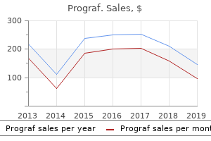
"Order prograf with paypal, hiv infection mouth ulcers".
By: U. Kasim, M.B. B.A.O., M.B.B.Ch., Ph.D.
Professor, Indiana University School of Medicine
In addition to its classic presentation in the diaphyses of long bones hiv infection rate in india discount 0.5mg prograf free shipping, the tumor can contain axial bones such as the pelvis and ribs hcv hiv co infection rates buy prograf master card, as properly as gentle tissues (see Chap antiviral yahoo generic 1 mg prograf amex. Although patients often have pain and a mass, they could additionally current with fever and leukocytosis suggestive of an infectious course of. Evidence of neuroectodermal differentiation could also be manifested by extracellular eosinophilic neuropillike constructions or Homer-Wright rosettes (composed of teams of tumor cells that encompass a central core of eosinophilic extracellular material). Most tumors current after 30 years of age with a peak incidence between 50 and 60 years of age. The midline of the axial skeleton, particularly the sacrum and the bottom of the skull, is usually affected. Chordomas are aggressive tumors that are most notable for local recurrence when incompletely excised, but additionally have a metastatic potential. A controversial entity, "chondroid chordoma," characterised by areas of mimicking hyaline cartilage, seems to have a greater prognosis, whereas "dedifferentiated chordoma," with its high-grade sarcomatous element, is related to a really poor end result. Simple bone cyst is an intraosseous space-occupying lesion consisting of an accumulation of fluid. The lesion is often lined by a skinny membrane composed of flattened cells of unknown type which may be involved within the manufacturing of the fluid, which usually seems serous. The base of the lesion is normally situated at an active progress plate, and the cyst is kind of maintained by continuous transforming of the bone across the space of fluid stress. Histologically, the diagnosis is considered one of exclusion and relies upon upon correlation of the imaging, operative findings, and lack of any other diagnostic tissue. The defect is histologically much like the subarticular cysts associated with overlying osteoarthritis (geodes) besides that the subarticular plate and articular cartilage are radiographically intact in patients with intraosseous ganglia. Within the septae are fibroblasts, scattered multinucleated large cells, and osteoblasts related to the bone manufacturing. Rarely, the lesion is nearly totally strong, although sometimes the solid variant might reveal fluid levels on imaging. The genetic abnormalities so far seem to apply primarily to the first number of this lesion, and have additionally been discovered in the solid variant (Am] Pathol. There are different benign and malignant soft-tissue neoplasms that can primarily come up in bone, including leiomyoma, leiomyosarcoma, lipoma, liposarcoma, and schwannoma. All of those are histologically just like their soft-tissue counterparts (see Chap. Because of its accessibility and composition of each cortical and trabecular (cancellous) bone, the iliac crest is the positioning of choice for evaluation of systemic metabolic bone diseases. Trabecular bone, the meshwork surrounded by marrow or fat, is much more metabolically active than cortex. To assist both its mechanical and metabolic functions, bone is dynamically regulated, and the skeleton is changed fully every 10 years. The web results of each cycle is the formation of a brand new osteon, a packet of bone delineated by a "cement line" in which the collagen fibers are aligned. However, the strength of the undecalcified biopsy is in the evaluation of the function of these cells rather than their morphology. The thickness of the osteoid seams displays the rate of mineral apposition, as a outcome of mineralization converts osteoid to *All e-figures are available on-line by way of the Solution Site Image Bank. Decalcification required for standard paraffin processing removes the excellence between newly synthesized osteoid and mature calcified bone. In distinction, undecalcified plastic sections may be stained in a number of methods to show osteoid. These latter stains permit easier interpretation of cellular morphology than the von Kossa, and also highlight peritrabecular or marrow fibrosis. Tetracycline household antibiotics are calcium-chelating fluorochromes that bind to actively mineralizing bone surfaces, may be taken orally, and are well-tolerated. Combination of the extent of labeled trabecular bone floor and the gap between labels provides the mineral apposition fee and bone formation price. In a standard topic, most surfaces with osteoid, as seen on trichrome or von Kossa stains, have single or double labels. Biopsy is performed on the third day after the last dose (biopsy interpretation could subsequently be confounded by recent therapeutic use of antibiotic medicine in the tetracycline family; equally, insufficient labeling could be attributable to malabsorption syndromes or by taking tetracycline with meals, dairy merchandise, iron-containing medicines, antacids, or calcium supplements).
Brushing samples can be smeared immediately on a slide anti viral hand gel generic prograf 5mg overnight delivery, however care have to be taken to repair smears instantly or air-dry artifact will render the slides uninterpretable antiviral movie order prograf in united states online. Brushes could also be mounted in fluid rate of hiv infection in jamaica purchase prograf on line, and cell block or Pap stained specimens may be produced from materials dislodged from the comb into the fluid. Fine needle aspirates of lung lots, whether or not obtained through endoscopic or transthoracic approaches, are DiffQuik and Papanicolaou stained. Pleural effusions are typically submitted contemporary, in toto, from which a well-mixed portion (generally 2-300 ml) is used to put together each a cytospin DiffQuik and ThinPrep Papanicolaou stained slide preparation. This diagnosis is rendered when the specimen shows only alveolar macrophages, benign bronchial epithelial cells, and mixed inflammatory cells. This prognosis can also be used when fungal elements are recognized, or when viral cytopathic modifications are seen. This analysis is rendered when rare malignant cells are present, however the amount is insufficient for a definitive analysis of malignancy. This prognosis usually prompts a repeat diagnostic procedure earlier than definitive surgical therapy. This analysis is rendered when each the quality and amount of malignant cells are enough for an unequivocal diagnosis of malignancy. Diagnoses of malignancy are greatest considered a operate of each high quality and amount of atypical cells; both rare markedly atypical cells or an abundance of only minimally atypical cells can recommend malignancy. The radiologic correlates to viral infection sometimes embody diffuse pulmonary infiltrates. Cytopathologically, viral changes are best appreciated on bronchial washing and bronchioloalveolar lavage specimens. The viral cytopathic changes seen in Herpes virus infections include multinucleate and single infected cells which have massive nuclei with clear to faintly basophilic centers and peripheralized chromatin; the nuclei are molded with one another. Adenovirus, which yields a typical "smudged" appearance on histologic part, shows a polygonal nuclear inclusion and multinucleate cells. The chest X-ray of sufferers with fungal infection can exhibit both a mass impact, a number of nodules, or, every so often, diffuse infiltrates. Alveolar casts containing fibrin and the organisms have a bubbly or foamy look on Papanicolaou-stained supplies. Methenamine silver stains can highlight the organism, and the stain can be utilized to formalinfixed paraffin embedded cell blocks, or to cytospins. Patients taking prophylactic antibiotics for Pneumocystis could not have alveolar casts, and their samples could have rare, cup formed organisms positioned within macrophages. In the latter, concretions of allergic kind mucin or Charcot Leyden crystals rna y be noted. Histoplasma infection not often shows detectable organisms; nevertheless, a granulomatous reaction may be current in aspirations of pulmonary masses or of concerned lymph nodes. Cryptococcus, Coccidiomycosis, and Blastomycosis can all induce solitary masses, which may be sampled by aspiration. The yeast forms are best appreciated on Romanowsky stains; typically the organisms are clear. Again, cell blocks, direct smears, or cytospins may be stained with traditional techniques to higher spotlight the organisms. The prototypical bacterial an infection for which cytologic sampling is pursued is tuberculosis. Washings or lavage may be pursued primarily for acquiring material for tradition, molecular laboratory check to detect the organism, or antibodymediated studies. Cytopathologically, washings or lavage fluid can show no important adjustments or could exhibit necrosis and acute irritation. Cavitary lesions can have secondary squamous metaplasia, which can be atypical; for this reason, the chance of a false constructive cytology should at all times be considered in the setting of a cavitary lung mass. Community acquired bacterial pneumonias are less usually sampled, however once more, fluids from such cases exhibit acute inflammation, macrophages, and respiratory epithelial cells. Sarcoidosis is a prognosis of exclusion, and fantastic needle aspiration from sufferers with infiltrates and adenopathy may be used as a minimally invasive method for evaluation. Aspiration of lymph nodes form patients with hilar adenopathy yields fragments of granulomas, rare multinucleate large cells, and a background of mixed continual inflammatory cells. The background contains mixed acute and chronic inflammatory cells, with variable numbers of eosinophils. Alveolar hemorrhage from any cause may find yourself in the presence of hemosiderin laden macrophages, highlighted by iron stain.
0.5mg prograf sale. AntiViral.
After weighing the complete specimen hiv infection wiki discount prograf 0.5 mg mastercard, the renal hilum is examined to identify the ureter anti viral hand foam norovirus purchase prograf 5mg visa, renal vein hiv infection impairs quizlet order prograf 1mg amex, and artery, and cross sections of those margins are taken. Sections via renal masses ought to demonstratt: relationship of mass to capsule, peripheral fats, renal parenchyma, and renal pelvis. Tumor dimension, location (upper or lower pole, cortex, or medulla), involvement of calyceal or pelvic mucosa, invasion of the capsule or perirenal delicate tissue, and involvement of the adrenal gland are recorded. The uninvolved parenchyma can be examined: color, cortical thickness, extra focal lesions, and renal pelvis are described. One part per centimeter of tumor, demonstrating its relationship to adjacent capsule, renal parenchyma, and pelvis are submitted, as properly as sections of any additional lesions, uninvolved renal parenchyma, and adrenal gland. Additional danger components include weight problems, hypertension, unopposed estrogens, and exposure to arsenic, asbestos, cadmium, organic solvents, pesticides, and fungal toxins. About 5% of sufferers with acquired cystic disease of the kidney develop renal cell tumors (see part on Acquired Cystic Disease-associated Renal Cell Carcinoma). Hereditary leiomyomatosis and renal cell cancer is an autosomal dominant illness caused by mutations in the fumarate hydratase gene. Tumors have papillary sort ll giant eosinophilic cells with large nuclei and nucleoli. Birt-Hogg-Dube syndrome is characterised by cutaneous fibrofolliculomas, trichodiscomas, and acrochordons. Several tumor varieties have been seen, together with clear cell carcinoma, papillary carcinoma, and oncocytoma. Hematuria, ache, and a flank mass is the classical triad of presenting symptoms, however in North America most renal tumors are now detected as incidental findings by radiological studies. Other shows embody weight reduction, anorexia, fever, hypercalcemia, erythrocytosis, hypertension, gynecomastia, anemia, and hepatosplenomegaly. Histologic sections show spherical or polygonal cells with clear or eosinophilic cytoplasm and centrally located nuclei. The tumor cells are typically of excessive nuclear grade and may have hobnail cytology. Tumors in adults are identical to those within the pediatric age group as mentioned earlier. Uncommonly, necrosis, clear cells papillations, foamy macrophages, and irritation could be current (Am] Surg Pathol. Features that place tumors in this group embody the next: a mix of various histologic sorts, mucin production, sarcomatoid appearance, presence of epithelial and stromal parts, and nonidentifiable patterns. Heterologous malignant bone, cartilage, fats, and skeletal muscle or homologous undifferentiated malignant spindle cells could be seen. Tubulocystic carcinoma is very unusual and is of low malignant potential (Am] Surg Pathol. Grossly, the tumor is often solitary and circumscribed with a spongy "bubblewrap"-like cut floor. Additional architectural patterns embody stable acinar, solid sheet-like, papillary, and macrocystic. The papillae are prominent and lined by cells with variable amounts of cleared cytoplasm. A attribute finding is linear arrangement of the nuclei in the middle or apical portions of the cytoplasm. Most tumors are low stage at presentation and limited consequence information recommend an especially favorable prognosis. Thyroid-like follicular carcinoma of the kidney is uncommon, with lower than ten instances reported (Am] Surg Pathol. Papillary adenoma of the kidney is the most typical benign epithelial neoplasm of renal cells. Grossly, the tumor consists of single or multiple welldefined white nodules within the renal cortex. The lesion is extra frequent in men, and the height incidence is through the seventh decade of life. Microscopic (but not gross) extension into perirenal fat and vessels has been described. Metanephric adenoma happens in children, and in adults through the fifth and sixth a long time.
Necrotizing (malignant) otitis externa is usually brought on by Pseudomonas aeruginosa infecting diabetic patients oral hiv infection symptoms purchase prograf 0.5mg fast delivery, but fungi can additionally be the causative agent hiv infection youth order prograf 5 mg fast delivery. The most typical neoplasms of the exterior ear are basal cell carcinoma and squamous cell carcinoma of the skin of the ear hiv rates of infection in us discount 1 mg prograf with visa. Malignant tumors of ceruminous glands embody adenocarcinoma, adenoid cystic carcinoma, and mucoepidermoid carcinoma. Ceruminous adenocarcinomas present infiltrative development and range from cytologically bland to markedly atypical with an increase in mitotic activity. Perineural invasion is rare, however could be a useful diagnostic clue favoring adenocarcinoma when detected, especially in small biopsy samples. Other uncommon neoplasms and tumorlike circumstances of the external ear embody malignant melanoma, benign fibro-osseous lesion, osteoma and exostosis, and idiopathic pseudocystic chondromalacia (which is a nonneoplastic swelling of the pinna because of fluid accumulation throughout the cartilage of the ear). The frequent problems embody choristoma, irritation, an infection, ldl cholesterol granuloma, cholesteatoma, and otosclerosis. Choristomas (heterotopic tissues) within the center ear are composed of benign salivary gland, glial, or sebaceous gland tissue. Most acute purulent instances are due to bacterial infection by Streptococcus pneumoniae or Haemophilus influenzae. Microscopically, granulation tissue, scar tissue, chronic irritation, calcific particles, and sclerotic or reactive bone may be seen. There may be associated polypoid granulation tissue, ldl cholesterol granulomas, cholesteatoma, or tympanosclerosis. The congenital type is found in infants and young youngsters, is defined as occurring in the presence of an intact tympanic membrane, and will end result from an epidermoid cell rest (epidermoid formation). Downgrowth of the epithelium into underlying subepidermal connective tissue could also be appreciated. There is an increased cell proliferation index in the squamous epithelium of cholesteatoma (Acta Otolaryngol. Otosclerosis is a disease of unknown etiology that results in progressive fixation of the stapes footplate and, in consequence, conductive listening to loss. Chapter 7 � the Ear I one hundred and one the initial histologic modifications embody resorption of bone and alternative by mobile fibrovascular tissue. Neoplasms of the center ear include paraganglioma, adenoma of the center ear, papillary tumors, meningioma, and squamous cell carcinoma (Table 7. Paraganglioma (also known as glomus tumor, glomus tympanicum, or chemodectoma) is the most common tumor of the center ear (but is nonetheless nonetheless rare). It usually presents clinically with hearing loss or tinnitus in patients in the fifth and sixth decades of life. Clinically, paraganglioma is bulging, purple, pink, or bluish (and not white like a cholesteatoma). Immunohistochemical stains can be confirmatory and particularly contributory in small biopsy samples, which can show important crush artifact. Chromogranin A and synaptophysin immunostains are optimistic, whereas carcinoembryogenic antigen and keratin immunostains are unfavorable. Intracranial extension develops in a small minority of patients, and about 1% to 2% of patients undergo from metastatic unfold. Adenoma of the center ear is a benign glandular neoplasm with variable neuroendocrine and mucin-secreting differentiation (Arch Pathol Lab Med. The typical clinical presentation is in an adult (mean age within the 40s) with muffled hearing, tinnitus, and/or a sensation of stress and/or fullness. The neoplasms are variably colored and solely uncommonly penetrate through the tympanic membrane. The immunoprofile consists of positivity for cytokeratin and neuroendocrine markers such as chromogranin and synaptophysin. Recurrence has been reported in a small minority of circumstances, often after incomplete surgical excision. Papillary tumors ofthe middle ear embrace aggressive papillary tumor, Schneiderian papilloma, and inverted papilloma, although only a few cases of the latter two neoplasms have been described.